Brain Scan Can Reveal How Much School Work You’ve Done
 Much like the age of a fish can be determined by the number of rings on its scales, a new study shows that the number of years a person has studied in school can be seen on a brain scan decades later by measuring the growth in certain brain regions. Moreover, the increased gray matter from hitting the books protects against cognitive decline in aging.
This new study, led by Gaël Chételat at INSERM in France, investigated both structural differences and functional differences in the brains of healthy elderly individuals. The subjects were prescreened to detect amyloid deposits in the brain to exclude people who might have preclinical Alzheimer’s disease. “This is important,” Dr. Chételat explained, “because it allows us to specifically address the relationship to education without the effect of pathology that complicates interpretation.”
The battery of tests included MRI brain imaging to pinpoint anatomical differences in the brain, PET imaging to assess metabolic activity in different regions of brain tissue, and functional MRI, which examines neural activity and functional connectivity in the brain at rest. The studies were performed in people between the ages of 60 and 80 who had from 7 to 20 years of school education.
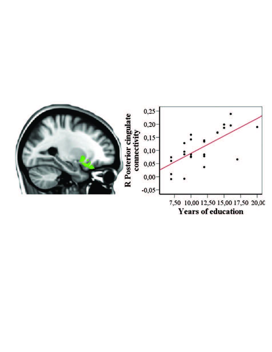
The findings showed a linear increase in gray matter volume in proportion to the number of years of education the elders had completed in their youth. This included increases in three brain regions (right superior temporal gyrus, anterior cingulate gyrus, and left insular cortex). Brain metabolism also increased proportionately with years of education in the anterior cingulate gyrus, and functional connectivity between several brain regions that are critical for cognitive function increased linearly with the number of years of education. This included increased connectivity with the anterior cingulate gyrus and the right hippocampus, left angular gyrus, right posterior cingulate, and left inferior frontal gyrus–all circuits known to be important in memory and cognition.
Thus, both the structure of the brain and its function in old age are increased in proportion to the number of years of education received decades earlier, and what’s more, these lasting fringe benefits of classroom work benefit healthy elderly people who are free of Alzheimer’s disease. The more years of schooling a person has, the more resistant people are to cognitive decline in aging because they have both more brain tissue in critical regions, and the level of metabolic function in their gray matter is elevated.
Epidemiological studies over the last decade have shown a lower incidence of dementia among individuals in the elderly population who have higher levels of education and also found that Alzheimer’s disease is delayed in this population. Prior to this new study it was unclear whether the increased brain metabolism, higher functional connectivity, and bulked up grey matter seen in aged individuals with increased education occur simultaneously or are independent effects, because these prior findings had been identified in separate studies. Also unclear was whether these benefits were granted by chance from birth, promoting a person’s success in school and staving off cognitive decline in later life because of the increased “cognitive reserve.” That is, people with more robust brains can withstand the erosion of mental capabilities during aging because the greater mental capacity allows them to resist the negative effects longer.
The authors conclude that while people with genetic advantages may excel in school, school work itself benefits everyone by building stronger and more active brains. “It is crucial to promote the beneficial effects of environmental factors: food, sport, mind and cognition,” Dr. Chételat says, as complementary approach to medication in resisting cognitive decline with age.
Arenaza-Urquijo, E.M. et al., (in press) Relationships between years of education and gray matter volume, metabolism and functional connectivity in healthy elders. NeuroImage, available on-line in advance of print doi: 10.1016/j.neuroimage.2013.06.053.