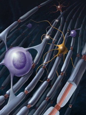
The Brain’s White Matter–Learning beyond Synapses
Neural computation and information processing take place in the brain’s gray matter, the “topsoil” layer of brain tissue comprised of neurons communicating through synapses, but beneath gray matter billions of nerve axons connect neurons into circuits, much like underground communication cables connecting computers into functional networks over long distances. These dense bundles of cables in white matter held little interest to neuroscientists who were interested in how the brain processes information and learns. These functions were understood to depend on the formation and breaking of synaptic connections between neurons as well as adjustments in the strength of synaptic connections. But as any audiophile knows, cabling is critical to the performance of any communication system. Moreover, while it is important to board a train efficiently–much like signals entering a neuron through its synapses–the time it takes to travel between destinations can be even more important in the optimal function of the network compared with the process of “crossing the gap” into the train car. This concept, that the time it takes to transmit signals over long distances between neurons in the brain is important, has been largely overlooked in neuroscience theories of learning.
The most important factor in determining the maximal rate of impulse transmission through a nerve axon is whether or not the axon is coated with electrical insulation called myelin. Myelin boosts the speed of electrical impulse transmission by at least 50 times. People have always understood that if the myelin sheath breaks down in disease, such as multiple sclerosis, impulses can fail to travel beyond the damaged insulation and the neural circuit fails. In multiple sclerosis, for example, failure of transmission because of myelin damage results in blindness or difficulty walking. But what about the possibility that myelin might influence the proper rate of transmission between relay points in a neural circuit by adjusting the transmission speed so that impulses, like people arriving at appropriate times to catch a connecting flight, arrive at the right time at critical points in the circuit?
Evidence for changes in white matter structure during learning has accumulated in recent years from human brain imaging studies. At the same time, cellular neuroscientists have found that the formation of myelin can be influenced by electrical activity in axons. These two clues suggest that myelin may change during learning to optimize the flow of information through neural circuits. In a study to be published in Friday’s edition of the journal Science, a team of researchers from the laboratory of William Richardson at University College London, and colleagues in Australia, Japan, and Portugal, provide new evidence that learning a motor skill requires new myelin to be formed in the brain.
In the brain and spinal cord, myelin insulation is wrapped around axons by glial cells called oligodendrocytes. These cells develop from immature glia called oligodendrocyte progenitor cells (OPCs). Curiously, these immature glia persist in the adult brain in great numbers. In fact, OPCs are the major class of dividing cells in the adult brain. They comprise 5% of all cells in your brain. Why are these immature glia present in the adult brain long after fetal development?
One possibility is that these immature glia are there waiting to form myelin on bare axons in the adult brain to boost the speed of transmission in a circuit that is engaged in learning a new skill. If so, this would be a non-synaptic mechanism of learning. To test this hypothesis, the scientists studied mice that had been genetically engineered to prevent the OPCs from maturing into oligodendrocytes. They were able to control this block of OPC maturation by giving the mice a drug (tamoxifen) to block this process at any point in the animal’s life. The question they asked is whether or not new myelin is needed to learn a new skill, and by blocking the ability of OPCs to mature into oligodendrocytes, the formation of new myelin would be impaired while leaving myelin on existing fibers intact.
The mice were trained on a running wheel that had some of the rungs missing. At first the mouse stumbled over these missing rungs, but with practice, the animal learned to anticipate the missing rungs and step past them nimbly to run the treadmill with ease. The results showed that the mice that could not make new myelin learned this task much slower than normal mice. So no matter what changes in synaptic communication were taking place in gray matter to help the mouse learn to run on the modified wheel, if new myelin could not form, the animal’s performance would be impaired.
The researchers found that when an animal is trained on the running wheel, OPCs started to divide in its brain (in the corpus callosum, which are axon cables connecting the right and left hemisphere). By 4-6 days after training, there was a 40% increase in the number of OPCs that were dividing, and by three weeks there were many newly formed oligodnedrocytes. When this increase in oligodendrocyte production was impaired by genetic manipulation, the ability of mice to learn to run on the wheel was impaired.
An interesting finding was that quite similar increases in OPC cell division were also seen in the brains of control mice that were allowed to run on a standard running wheel, compared with mice kept in a normal cage without a running wheel. Possibly motor functions common to wheel running, such as grasping the rungs, are improved through myelination by running on the normal wheels as well. Alternatively it might be that exercise or novelty increase OPC production in the adult brain, not necessarily learning specifically. This benefit of enriched environments and exercise on OPC production in the adult brain is something that has been noticed in several other studies. This raises the question of whether new myelin is formed by the learning process to speed impulse transmission through the circuit required for the task, or instead there might be a general benefit of capacity for myelination in the adult brain by providing a better network structure for motor learning and performance.
Studies in aged humans show that myelin begins to degrade in aging and that learning new skills improves the integrity of white matter in the brain by slowing loss of myelin (Engvig, et al., (2012). The mice in this study were not aged, but still there might be a constant process of myelin renewal that could be difficult to detect but important for motor performance. This renewal would be undermined by the genetic manipulation blocking the formation of new myelin. In an e-mail, William Richardson agrees that this is an alternative possibility, but he doubts that this is as important as the formation of new myelin on circuits that must improve their performance during motor learning. “There is no detectable loss of oligodendrocytes after one month or three months,” the age at which these studies were performed, he says.
Richardson also offered a peek at what may be on the horizon. In comparing the rates of learning between controls and mice unable to form new myelin they noticed, “That the difference between [experimental and control mice] starts to develop less than 2 hours after first introduction to the wheel. At face value, therefore, myelin…is involved in the early events as well as the later events [of learning].”
This is intriguing because recent brain imaging studies have detected changes in white matter structure in the human brain by magnetic resonance imaging within two hours of training on a race car video game (Hofstetter et al., 2013). “Maybe the early events involve new protein production (Wake et al. 2011), while the later events contribute to long-term memory/consolidation,” Richardson says. (The study he is referring to found that the synthesis of myelin proteins is stimulated by electrical activity in axons, so that oligodendrocytes in contact with axons firing impulses begin to form myelin rapidly on those fibers.)
After a century of research focused on the synapse to understand the cellular basis of learning and memory, neuroscientists are excited to find that there may be much more in store in the neglected white matter regions of the brain involving non-neuronal cells, glia, that have been largely overlooked by most neuroscientists. The questions presented by this elegant new research are fascinating. For example, how do oligodendrocytes know which neural circuit to myelinate during learning, that is, how do they sense electrical activity in axons? What are the molecular mechanisms that control activity-dependent myelination? If these molecules can be identified, new approaches to treating nervous system disorders might be found, because abnormal transmission of information is associated with mental illnesses as well as neurological illnesses. How is this cellular mechanism of learning different from learning based on synaptic plasticity? For example, do improvements in transmission of information through a neural network explain why learning a difficult motor skill, such as riding a bike, takes so much practice, but suddenly everything kicks in and once the training wheels come off, you never forget how to ride a bike the rest of your life?
“Plenty more experiments up ahead!” Richardson exclaims.
More to explore
Bracken, Kassie (2009) The brains behind talent, The New York Times http://www.nytimes.com/video/sports/1194817108368/the-brains-behind-talent.html
Engvig, A., et al., (2012) Memory training impacts short-term changes in aging white matter: a longitudinal diffusion tensor imaging study. Human Brain Mapping 33, 2390-2406.
Fields, R.D. (2008) White Matter Matters, Scientific American (March 2008) 298, 54-61.
http://www.nature.com/scientificamerican/journal/v298/n3/full/scientificamerican0308-54.html
Fields, R.D. (2012) Social interaction in early life affects wiring to the frontal lobes The Huffington Post November 13, 2012
http://www.huffingtonpost.com/dr-douglas-fields/social-interaction-early-life-frontal-lobes_b_1864234.html
Hofstetter, S., Tavor, I., Moryosef, S.T., and Assaf, Y. (2013) Short-term learning induces white matter plasticity in the fornix. J. Neurosci. 33, 12844-50 (2013).
**McKenzie, I., Ohayon, D., Li, H., Paes de Faria, JH., Emery, B., Tohyama, K., and Richardson, W.D. (2014) Motor skill learning requires central myelination. Science, October 17, 2014 issue.
Wake, H., Lee, P.R. and Fields, R.D. (2011) Control of local protein synthesis and initial events in myelination by action potentials. Science 333, 1647-51.
** Reviewed in this article.
