To Flee or Freeze? Neural Circuits of Threat Detection Identified
 Suddenly something streaks into your peripheral vision. Instantly, you jump back and raise your arms defensively. “What was that!” You exclaim in shock. Only then do you realize that the blurred streak you just dodged was a wayward basketball zinging like a missile on a collision course for your face. A rush of adrenaline flushes through your blood setting your heart pounding and muscles twitching, but there is nothing left to do. Your brain’s rapid response defense system has already detected the threat and avoided it before your conscious mind is even engaged. How is that possible, scientist, Peng Cao and colleagues of the Chinese Academy of Sciences wondered?
The mystery runs deeper. The sight of a sudden threat can trigger the completely opposite response–you may freeze like a deer in the headlights. Sometimes freezing is the best move. Spotting a rattlesnake in the bush, perhaps, is best handled by freezing. Running away could provoke the reptile to strike. But neither response–freezing nor fleeing–is a deliberate, conscious reaction to a threat that looms so quickly you can scarcely perceive what it is. These familiar facts must mean that all of the neural processing for this life-saving reaction takes place in neural circuits that are not located in the cerebral cortex where consciousness arises. These neural circuits that suddenly grip control of your behavior, known as the “fight-or-flight” response, must reside in deeper layers of the brain.
Previous research has shown that there is a high-speed pathway from the retinas in our eyes to the center of the brain’s threat detection region, which includes the amygdala and related structures comprising the limbic system. The majority of visual information detected by the retinas is transmitted to the cerebral cortex at the back of the brain, where complex analysis enables us to interpret the shifting patterns of light and shows cast on our retinas as objects in space, with color, dimension, motion, and identity. This sophisticated visual processing takes time. The subcortical pathway from the eyes to the amygdala is fast, but we are not able to actually see what the object is, because the necessary analysis for vision requires the cerebral cortex. But that route through the visual cortex takes far too long to dodge something like an opponent’s right hook. This high-speed, subcortical threat detection pathway is like a motion detector in a home security system. A moving object in the environment sets off an alarm that there is an intruder. Whatever it is, we can’t say for sure, but it should not be there!
Cao and colleagues traced out this circuitry in detail and they have identified the specific neurons that control whether we flee or freeze when an object suddenly looms in our visual field. The first relay point for high-speed information transmission from the retina to the brain is a region called the superior colliculus. There are three different types of neurons in the superior colliculus that can be identified by the different types of proteins that are contained in them. One set of neurons contains a protein called parvalbumin (PV). Mixed in with these, are neurons that contain either the protein somatostatin (SST) or vasoactive intestinal peptide (VIP). The researchers found that when they stimulated the PV neurons, the mouse immediately bolted or froze.
To stimulate these neurons selectively, the researchers used genetic manipulation to insert light-sensitive ion channels specifically into the PV neurons. These channels will activate when stimulated by light delivered through a fiber optic cable surgically implanted into the mouse’s brain, thereby causing the PV neurons to fire electrical impulses. When researchers flipped on the fiber optic light, the mouse fled away, and then it cowered after they stopped stimulating the PV neurons. This suggests that the PV neurons are a vital part of a threat detection circuit in the visual pathway. This function of PV neurons was further supported by monitoring the electrical activity in these neurons in anesthetized mice. The researchers found that when a virtual object on a computer screen that resembled a soccer ball came flying directly toward the animal’s head, the PV neurons began firing electrical impulses vigorously. The mouse’s heart rate accelerated and the stress hormone corticosterone increased in the blood stream–the bodily responses we experience as fear in the fight-or-flight reaction. But if the ball moved through the visual field in any other direction except on a collision course, the PV neurons remained silent. The mouse’s heartrate remained calm.
But what determines whether the animal flees or freezes? Interestingly the researchers found that the same neurons controlled both behaviors. Strong stimulation of the PV neurons caused the animal to escape rather than freeze. Either a brighter laser beam, or longer pulses of light, or higher frequency of flashes, would cause the animal to escape rather than freeze.
An interesting observation was that male and female mice responded somewhat differently. Females tended to escape, whereas males tended to freeze–stand their ground, perhaps, in the face of a sudden visual threat that stimulated these PV neurons. Further research will be required to uncover the additional factors that predispose males and females to respond differently to the same visual threat. The researchers then traced the circuit from these neurons and found that they did indeed connect to the amygdala, via a relay neuron in a part of the brain called the PBGN (parabigeminal nucleus). Further analysis showed that PV neurons stimulated neurons to fire by using the excitatory neurotransmitter glutamate. This is unusual because PV neurons elsewhere in the brain use a different neurotransmitter (GABA) to inhibit firing of the neurons they connect to.
This work advances our understanding of how visual threats trigger a fight-or-flight response, but there is much more to be discovered. “What are the functions of the other two pathways?” Peng Cao asks in response to my question about the next step in his research. (He is referring to the function of the SST and VIP neurons in the superior colliculus.)
“Do human beings share a similar pathway with rodents?” He wonders. Cao’s hunch is that these neurons are relevant to fear disorders. “We speculate that this pathway in mice may be genetically defined and subject to environmental modifications.” If humans have the same circuitry from their retina to the amygdala via PV neurons in the superior colliculus, Cao suspects that, “this pathway may be involved in fear disorders such as PTSD.” The amygdala is involved in fear and in learning to avoid dangers, but in addition to this anatomical evidence suggesting that PV neurons may be involved in fear disorders, Cao and his colleagues noticed something interesting. When they stimulated this pathway in the superior colliculus of mice repeatedly, the mice began to show depression and avoidance-like behaviors, much as people do who develop PTSD after surviving an extremely traumatic event.
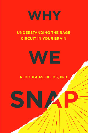
Note: Readers who are interested in this subject may be interested in my new book Why We Snap, to be published this year by Dutton and available for pre-order now. The unconscious neural circuitry of the fight-or-flight response is involved in many other responses to threats, fear, and when they misfire: snapping in rage.
http://www.penguinrandomhouse.com/books/316682/why-we-snap-by-r-douglas-fields/
Reference
Shang, C., et al., (2015) A parvalbumin-positive excitatory visual pathway to trigger fear responses in mice. Today’s edition of Science, June 26, 2015.
Photo credit: https://filmjamblog.wordpress.com/2012/11/11/the-light-house-cinema-book-club-the-shining/
