Neurological Implications of COVID-19 Raise Concerns
 Virologists are alerting doctors to a possibility that could help explain two of the most puzzling aspects of COVID-19—why the severity of the disease varies so widely, and how the infection can be so deadly. In severe cases the virus may enter the brain through the olfactory nerve in the nasal cavity and damage neurons that control breathing.
“Doctors should be aware of this possibility,” warns Pierre Talbot, Professor of Virology at the Institut Armand-Frappier, Université du Québec, Laval, Québec. “It may not be only pneumonia [killing patients]—the virus could be infecting the brain,” he says.
This is the conclusion reached in a new paper by Yan-Chao Li, Wan-Zhu Bai, and Tsutomo Kashikawa from the Jilian University of China and RIKEN Brain Science. According to the authors, patients suffering from COVID-19 who were admitted to intensive care in China could not breathe on their own, and many showed neurological signs, such as headache, nausea, and vomiting. Nearly all of the patients in ICU (89%) could not breathe spontaneously, and about half of the patients in intensive care worsened in a short period of time and died from respiratory failure.
Talbot concurs with the conclusions of those authors that neurons controlling breathing could be infected. “This is credible, and it is a major observation,” he says.
Microbiologist Stanley Pearlman, at the University of Iowa, who is an expert on the mode of infection of coronaviruses, agrees. “It is certainly plausible that this could be happening in a fraction of COVID-19 patients,” he says. “Maybe the ones with greatest severity of illness.”
For the last 25 years, Talbot and his colleagues have studied how coronaviruses infect the brain through the olfactory nerve. “Given our results with the human coronavirus causing common cold, I think we should not discard this possibility with the virus that causes COVID-19,” he says.
The coronavirus causing COVID-19 was unknown until a few months ago, so there is little information available, and the clinical picture of the disease is still being characterized. The human coronavirus causing up to 30% of common colds is genetically similar to the coronavirus causing COVID-19, Talbot says, but it is well established that many types of viruses can travel through the olfactory nerve into the brain. The list includes influenza A, herpes, West Nile, chikungunya, and most notably, SARS-CoV, the most closely related virus to the novel coronavirus that causes COVID-19. In fact, SARS-CoV, which causes SARS, shares 79% genetic identity with SARS-CoV2, the virus that causes COVID-19. Both viruses attack cells by binding to the same cellular receptor (ACE2), and they cause similar disease symptoms, including respiratory distress, fever, and highly lethal pneumonia.
Autopsy studies of patients who died in previous outbreaks of the closely related coronaviruses causing SARS and MERS found SARS-CoV virus in patients’ brains. Once inside the brain, the virus resides almost exclusively inside neurons rather than in other types of brain cells (glia), suggesting a trans-neuronal mode of infection rather than invasion of the brain from system-wide infection and entry through the blood-brain barrier, which can also happen.
Evidence is emerging that brain infection can also occur in COVID-19. Xinhua, the Chinese news agency, reported in early March on a 56-year-old patient in a Beijing hospital who had neurological symptoms that physicians described as “impaired consciousness.” Inspection of the patient’s cerebral spinal fluid led to a diagnosis of viral encephalitis, a type of brain inflammation that physicians attributed to the presence there of the COVID-19 virus. A non-peer-reviewed research report, posted Feb. 25 on the pre-print server medRxiv reports neurological symptoms in 78 severe cases of COVID-19 among a group of 214 patients treated at three Wuhan hospitals.
Such neurological symptoms can result from systemic disease, but coronaviruses can infect neurons. The incidence of neuronal infection in COVID-19 is unclear, because olfactory and brain tissue are not typically sampled at autopsy. The disease primarily attacks the lungs, but the cases involving neuronal infection would be the direst. Although neurons can be infected, the route of brain infection taken by coronaviruses is not known in people. Pearlman explains that by the time a patient dies and an autopsy is performed, the path the virus took to enter the brain is obscured by the widespread infection throughout the body.
Experimental research on the mode of SARS and COVID-19 infection cannot be performed in humans for ethical reasons, and studies cannot be performed on animals in laboratory research that do not have the human ACE2 receptor. To overcome this problem, Pearlman and colleagues genetically engineered mice to express the human receptor. His experiments on SARS-CoV published in 2008 showed that when the virus is delivered through the nasal cavity to mice that are genetically modified to express the human ACE2 receptor, it spreads to the brain through the olfactory nerves. Furthermore, the SARS virus was lethal when injected directly into mice’s brains, even though this mode of infection did not cause lung infection and pneumonia. Respiratory failure in this case is attributed to infection of neurons in the brain controlling breathing.
Recent reports that loss of the sense of smell can be an early symptom of COVID-19 provide additional circumstantial evidence of viral infection of the olfactory nerve and brain. “The lack of smell is often the first symptom of damage to the CNS,” Talbot notes. “It can be the first symptom of diseases such as Parkinson’s and Alzheimer’s, so it suggests that viruses that can get to the brain and could cause a loss of smell as a consequence of the virus traveling through the olfactory nerve.” He cautions, however, that such reports are anecdotal for now, but this should be monitored closely.
“There is already good evidence from South Korea, China and Italy that significant numbers of patients with proven COVID-19 infection have developed anosmia/hyposmia (loss or diminished sense of smell),” write the heads of the British Rhinological Society and ENT UK, which represents ear, nose and throat physicians there.
“In Germany it is reported that more than two in three confirmed cases have anosmia. In South Korea, where testing has been more widespread, 30 percent of patients testing positive have had anosmia as their major presenting symptom in otherwise mild cases,” continue the officials with the British organization. The American Academy of Otolaryngology has urged that loss of smell and related symptoms be added to the diagnostic criteria for COVID-19.
On the other hand, nasal infections frequently cause anosmia by affecting the sensory receptors in the nasal cavity, rather than the brain. In most cases, the normal sense of smell returns after a respiratory infection within about 14 days. Damage to the olfactory nerve or brain, however, can cause longer-lasting or permanent deficits. Talbot’s studies in mice on human coronavirus causing common cold show that infection of the sensory receptors and olfactory nerve can occur together, and that there are multiple ways that coronavirus can infect the olfactory nerve.
Whether neurons have the ACH2 receptor is currently a matter of debate, Talbot says, but his studies show that the olfactory nerve and brain can be infected through what is called “receptor independent transmission,” and this is the current focus of research in his laboratory. The mechanism is still unclear, but he suggests that “endocytosis is a way for the virus to get into the cell so it may not require the receptor.” (Endocytosis is a process by which cells engulf various substances, including particles and microbes.)
Important implications
Lung infection is the primary mode of COVID-19 disease, but neural infection by coronavirus can occur. The number of such cases in COVID-19 is unknown, but with so many people being infected, many cases would be expected, and these would be the most severe ones. The possibility that brain infection may be contributing to breathing failure in severe cases of COVID-19 has important implications. Respirators and other medical resources are severely strained in the current pandemic, forcing doctors in Italy, for example, to make heart-breaking decisions on which patients have priority if there are not enough resources.
Talbot says that doctors need to know about the possibility of brain infection in COVID-19, but he adds, “It is difficult to imagine that we could solve this problem of respiratory failure from CNS infection.” People are normally put on respirators to sustain them until pneumonia or other breathing problems can be treated, but does it make sense to do so if the disease has destroyed their ability to breathe and they will not survive? U.S. doctors are finding that some patients who survive COVID-19 cannot be taken off respirators.
Another consequence of neuronal infection raises future concerns for patients who recover from acute illnesses. The pandemic has hit so suddenly, possible long-term consequences of the infection are unknown. It is unclear whether the loss of sense of smell is permanent, for example, but recent studies report that the virus remains detectable in some cases for weeks after patients recover and in one patient for as long as 37 days post-recovery. Other viruses that infect neurons can persist for life, such as the herpes virus (HSV-1) that causes cold sores and the chickenpox virus (varicella) that causes shingles. This is because most neurons, unlike most other cells, are not replaced throughout life. So too may SARS-CoV2 reside inside neurons long after the initial lung infection and the pandemic are over.
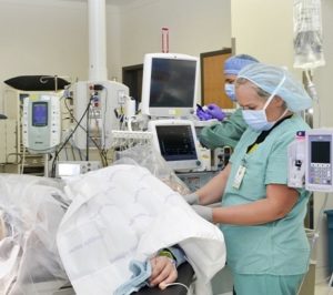
Photo credit: U.S. Army photo by Patricia Deal
This article first appeared in Psychology Today
I’m concerned that lipid nanotech coating of mRNA for Spine protein will directly pass though the blood brain barrier.
Spike protein’s kinetic structure- if activated by binding with any myelin protein is potentially destructive for vast white matter myelinated structures.
And myelin protein neuroantigen effects have been found to cross-react with Covid.
Spike protein dissolves into the myelin sheath – a huge volume of mobile gel structure. The trimeric structure of Spike suggests LEGO-like complexes might form when myelin and oligodendrocyte cells collapse.
The plaque-like hardness comes on quickly in lungs and brain and is uniform over nearly the entire surface.
As an osteopathic-trained child psychiatrist working with minocycline as an anti-inflammatory and clemastine as a stimulant for remyelination … I’ve been able to track the decrease in inflammation and the increase in white matter structures’ volume and elasticity via osteopathic palpation … and develop manual therapy to make sure remyelination is well-distributed throughout the brain.
We see relatively quick improvement in emotional self-regulation and learning disabilities – even communication in children with autism.
In Covid-19 acute illness and post-infection syndrome bilateral frontal lobe hardness matches bilateral lung hardness. Frontal lobe hardness does not release until lungs, liver, pericardium and heart release.
I suspect the reservoir of spike protein and the adhesions spike protein creates might be the driver for dysfunction. Recovery of lung function and Mental status improvement takes only 60-120 minutes of treatment.
I am very concerned about Moderna’s use of lipid-coated Spike mRNA.
Olfactory transmission through cranial nerve I is – by virtue of the mathematics of flow related to cross-sectional area- less than 0.1% of flow through myelinated fiber bundles.
White matter structures collapse readily. The shock of free passage through the blood brain barrier may resemble Zika virus effects more than SARS CoV-1 or 2.